Fascial Layers, Part 2 + Anatomy of a Nerve

For the most part, fascia can be classified as either superficial or deep, with the superficial layer being just beneath the skin and the deep layer being, well, everything else. As far as the deep layer goes, the “everything else” can be classified as either meningeal fascia or visceral fascia. 

DEEP FASCIA

Joint capsules, ligaments, tendons and the three layers that weave their way through each muscle (epimysium, perimysium and endomysium) make up the deep fascia. Aponeuroses are included in this classification as well. An aponeurosis is a broad, thick sheet of connective tissue that serves as an attachment site for muscles. A big one is the diamond-shaped thoracolumbar aponeurosis (where the latissimus dorsi originates) that spans across the lower back. I explained in Part 1 that fascia is rich in lots of sensory receptors. I’ll go into detail about each of those in a later post, but one of the sensory receptors found in fascia is called a nociceptor, otherwise known as a pain receptor. I once came across a study showing that people with lower back pain had a thickening of the thoracolumbar fascia compared to those in the trial who didn’t suffer from any LBP. This makes sense because with pain receptors being spread all throughout the fascial layers, a thickening of the fascia would equal an increase in pain receptors.

VISCERAL FASCIA

The visceral fascia is the layer that surrounds the heart (specifically called the pericardium) and lungs (pleura) as well as the abdominal organs. It also suspends the organs within their respective cavities (thoracic or abdominal) by way of ligaments that are meant to hold the organs against the body wall as well as allow for necessary physiological movement like breathing, the heart beating and peristalsis. 

MENINGEAL FASCIA

This is the layer that surrounds the brain and the nervous system. To better understand this, we’ll look at the anatomy of a nerve. But first...

WHAT IS A NERVE?

A nerve is a structured pathway that allows for the transmission of impulses to and from the brain and nervous system. Nerves either have one type of neuron, in which case they are classified as sensory or motor nerves, or like most nerves they have both motor and sensory neurons and are called mixed nerves. Nerves are structurally very similar to skeletal muscle in that each nerve has three separate layers of fascia, just like each muscle. 

Let’s look at the structure of a nerve from superficial to deep. The outer fascial covering of a nerve is called the epineurium (translates to on the nerve). Inside of that, nerve fibers (also called axons) are bundled together the same way muscle fibers are bundled, the layer that surrounds each bundle of axons is called the perineurium (around the nerve). Each individual axon that makes up the bundle is also surrounded by its own layer of fascia, this is called the endoneurium (within the nerve). 

Unknown-1.jpeg

Fascial Layers, Part 1 + Anatomy of a Muscle

While it’s true that the fascia is one big continuous and completely connected piece of tissue, it looks and acts differently depending on where it’s located in the body. In order to gain a better understanding of the fascial system as a whole and also have it be less overwhelming, we’ll break it down into more digestible bites. 

Just beneath your skin there is a layer of fatty tissue that provides the body with necessary insulation, blood and lymphatic flow and energy storage. Just beneath that is the superficial fascia. It anchors the skin to the tissues and organs below and is rich in blood and lymphatic vessels, nerves and some general sensory receptors which I’ll describe in detail in a later post. This thin and fibrous but highly elastic layer is classified as loose connective tissue. In this case loose just means it lacks any regular pattern or strong organization. 

Unlike the superficial fascia, the deep fascia is dense and well-organized. As far as I’m concerned, it’s the coolest layer of fascia because of the incredible and stunningly beautiful way it surrounds, supports and separates yet also connects every single structure in your body. The deep fascia is rich in sensory receptors that are sensitive to things like pressure and movement, which I will also cover in detail in another post. First let’s look at the anatomy of a muscle. Understanding the fascial anatomy of a muscle is essential for truly understanding how yoga and massage create change for people and actually really “work”.

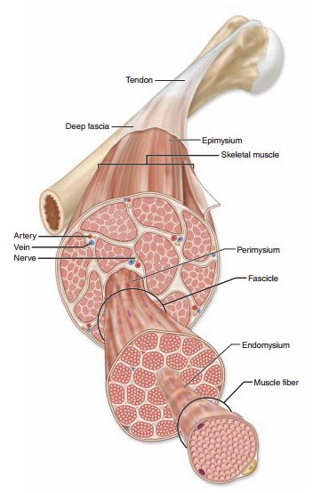
Let’s consider a muscle from the outside in, or anatomically speaking, superficial to deep. Every muscle as a whole is wrapped in a sleeve of fascia called the epimysium. Epi- meaning on and my- meaning muscle. Epimysium means on the muscle.

Within each muscle are groups of muscle cells that have been bundled together into what’s called fascicles. Each fascicle is wrapped in its own layer of fascia called the perimysium. Peri- meaning around and my- meaning muscle. Perimysium means around the muscle. 

Each individual muscle cell also has its own layer of fascia called the endomysium. Endo- meaning within, my- meaning muscle. Endomysium meaning within the muscle. 

Each of these three layers comes together to form the tendons that connect muscle to bone. The layer of fascia that surrounds each bone is called the periosteum.

In part two we’ll look at the fascial layers that surround the brain, nerves and organs as well as the anatomy of a nerve.

Unknown.jpeg

Collagen + Mechanical Properties of Fascia

Fascia has several mechanical properties that dictate how it functions. The main three are thixotropy, piezoelectricity and viscoelasticity.

Thixotropy refers to the ability of fascia to fluctuate between a gel (viscous) state and a sol (fluid) state, and it’s because of the ground substance that this can happen. Fascia is everywhere, and fascia has more ground substance than other types of connective tissue so while you’ve heard that we are made up of mostly water (about 70%), here is exactly what that means. The extracellular matrix (all the space outside of your cells in your connective tissue) is made up of about 90% water, and suspended in that water are a bunch of water-loving peptides called glycosoaminoglycans (GAGs). The GAGs attract water to keep the ground substance fluid so it can serve its purpose as a lubricant for the connective tissue. When connective tissue becomes dehydrated or injured, the ground substance loses fluid, causing fibers to stick together rather than slide and glide. We experience these areas called adhesions in our bodies as tension or knots. Hydrogen bonds the collagen fibers together, so when we release an adhesion through myofascial release or massage therapy, it’s the breaking down of the hydrogen bonds that can create that burning sensation we sometimes feel.

Let’s talk more now about how any of this applies to yin yoga and massage therapy. Piezoelectricity is another of fascia’s mechanical properties. It is derived from the Greek word ‘piezein’, meaning pressure or to squeeze. When subjected to gradual, sustained pressure, connective tissue produces a small electrical current across its surface. The current stimulates fibroblasts, the cells that produce fiber and ground substance. Pressure and movement increase the piezoelectric properties of fascia which in turn stimulate the healing process and contribute to the soft and loose feeling we often experience after a yoga class or a massage. This is why I often include elements of self massage and myofascial release in my classes. The third main property of fascia is the reason why that soft, loose feeling eventually goes away and ultimately why slow and steady wins the race when it comes to to reducing adhesions/pain and increasing flexibility, and why frequency matters more than duration.

In my classes I encourage the use of lots of props and for students to back away from their edge in their stretch. The reason is viscoelasticity: connective tissue’s ability to extend and then rebound rather than stretch and recoil. I mentioned in my introduction to fascia and connective tissue that collagen fibers have the ability to lengthen but they aren’t elastic so they can’t stretch without also sustaining structural damage. How is that?

The molecular component of a collagen fiber is called tropocollagen. Several tropocollagen fibers wind together and arrange themselves in a parallel alignment to form one collagen fibril, then multiple collagen fibrils wind together, also in parallel alignment, to form one collagen fiber. The fibers are all arranged into a triple helix which gives the fascia a great deal of tensile strength, meaning for the most part you can stretch it without it breaking. Collagen fibers do not stretch, but they do lengthen. When gradual, sustained pressure is applied, the collagen fiber unravels from its triple helix shape and eventually reaches its full length. This process of unwinding and extending can only happen when the force is applied gradually and repeatedly. When the force is sudden or extreme (such as bouncing in a stretch or going so deep into a stretch that your muscles are shaking), the collagen fibers resist and become even more bound. When the pressure is gradual and mindful, fibroblasts are stimulated to produce more tropocollagen, which forms new collagen and adds to the resting length of the existing fiber. This is why I teach so slowly and with so many props, it’s for the greater good of the fascial
system as a whole.

Sometimes almost as quickly as the release comes, we lose it. If you’re looking for increased flexibility, a decrease in pain, improved immunity, better sleep, a clearer mind and lots of other great things... stretch slowly and often, breathe deeply and give yourself permission to generally slow down. Keep your tissues hydrated by drinking lots of water and moving your body often, make the time to go to yoga, spend the money on the massage. The fascia is our internal environment and caring for it properly is essential for our overall health, happiness and wellbeing.