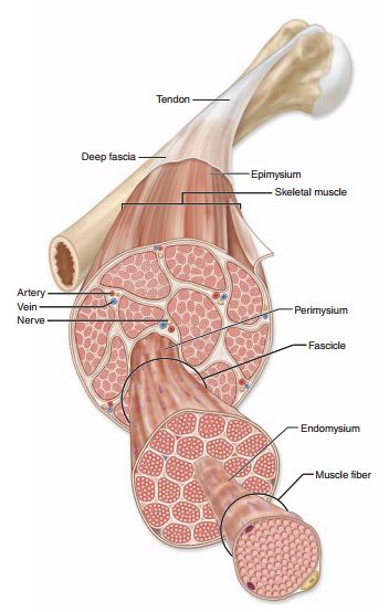
For the most part, fascia can be classified as either superficial or deep, with the superficial layer being just beneath the skin and the deep layer being, well, everything else. As far as the deep layer goes, the “everything else” can be classified as either meningeal fascia or visceral fascia.
DEEP FASCIA
Joint capsules, ligaments, tendons and the three layers that weave their way through each muscle (epimysium, perimysium and endomysium) make up the deep fascia. Aponeuroses are included in this classification as well. An aponeurosis is a broad, thick sheet of connective tissue that serves as an attachment site for muscles. A big one is the diamond-shaped thoracolumbar aponeurosis (where the latissimus dorsi originates) that spans across the lower back. I explained in Part 1 that fascia is rich in lots of sensory receptors. I’ll go into detail about each of those in a later post, but one of the sensory receptors found in fascia is called a nociceptor, otherwise known as a pain receptor. I once came across a study showing that people with lower back pain had a thickening of the thoracolumbar fascia compared to those in the trial who didn’t suffer from any LBP. This makes sense because with pain receptors being spread all throughout the fascial layers, a thickening of the fascia would equal an increase in pain receptors.
VISCERAL FASCIA
The visceral fascia is the layer that surrounds the heart (specifically called the pericardium) and lungs (pleura) as well as the abdominal organs. It also suspends the organs within their respective cavities (thoracic or abdominal) by way of ligaments that are meant to hold the organs against the body wall as well as allow for necessary physiological movement like breathing, the heart beating and peristalsis.
MENINGEAL FASCIA
This is the layer that surrounds the brain and the nervous system. To better understand this, we’ll look at the anatomy of a nerve. But first...
WHAT IS A NERVE?
A nerve is a structured pathway that allows for the transmission of impulses to and from the brain and nervous system. Nerves either have one type of neuron, in which case they are classified as sensory or motor nerves, or like most nerves they have both motor and sensory neurons and are called mixed nerves. Nerves are structurally very similar to skeletal muscle in that each nerve has three separate layers of fascia, just like each muscle.
Let’s look at the structure of a nerve from superficial to deep. The outer fascial covering of a nerve is called the epineurium (translates to on the nerve). Inside of that, nerve fibers (also called axons) are bundled together the same way muscle fibers are bundled, the layer that surrounds each bundle of axons is called the perineurium (around the nerve). Each individual axon that makes up the bundle is also surrounded by its own layer of fascia, this is called the endoneurium (within the nerve).

