Origin vs. Insertion

When talking about muscular attachments, it’s important to know and understand the difference between the origin and insertion. They are not interchangeable and have totally different meanings, though you can say muscular attachment or attachment site and be talking about either the origin or the insertion. 

What’s the difference? 
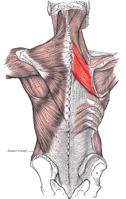
When a muscle contracts, the origin pulls the insertion closer.  Always! Muscles pull. The origin is the fixed point that doesn’t move during contraction, while the insertion does move. Your bones are the levers and your muscles are the pulley. Basically, we are all super complex puppets on strings. To explore this further, let’s take a look at the rhomboids. 

Rhomboids Minor and Major

Rhomboids Minor and Major

Firstly, there are two rhomboids and they’re both deep to the multidirectional fibers of the trapezius. Rhomboid minor sits on top of, or superior to, its larger counterpart rhomboid major. Minor originates on the spinous processes of C7 and T1 while major originates on the spinous processes of T2-T5. You can pretty easily feel for T1 on yourself, it’s just at the base of the posterior neck and it bumps out a little bit posteriorly. The transverse processes stick out to the sides while the spinous processes, which we are talking about here, stick out and a bit downward posteriorly. They remind me of a stegosaurus. It’s from these little posterior projections that both rhomboids originate. 

Back to rhomboid minor, it inserts on the upper portion of the medial border of the scapula, across from the spine of the scapula. The insertion for levator scapula is right there too and is easily palpable on yourself or someone else. Rhomboid major inserts on the medial border of the scapula as well, basically between the spine of the scapula and the inferior angle of the scapula. The actions of the rhomboids are to retract or adduct the scapula, elevate the scapula and also downwardly rotate the scapula.

WHOA, WAIT.
How does a muscle that elevates the scapula downwardly rotate it as well? If you look at the direction the fibers run, they’re on a bit of a downward angle going from the spine to the scapula. Because of this, when the muscles contract, they have the pulling capacity to draw the shoulder blades closer together. Downward rotation can be a tricky one because the scapula is the only bone in the body to do it, but we’re referring more specifically to the movement of the acromion, which is the flat bony process at the lateral end of the posterior scapula. When the rhomboids contract, they have the ability to tilt the acromion downward. It’s a slight action but it’s there nonetheless. Lastly the rhomboids can also elevate the scapula as a whole. Every time the muscle contracts (concentrically, that is, but that’s another post for another day), the origin says ‘come to me’, and so the insertion does. This is true for all the muscles in the body.

When studying anatomy, learn to look closely at the direction of the muscle fibers, and determine which end is the origin and which end is the insertion. That information will take you a long way in figuring out what actions a muscle can do.

ANOTHER EXAMPLE
Let’s look at a neck muscle- the sternocleidomastoid, or SCM. It has two heads which means it originates in two spots: the top of the manubrium (sternum) and the medial third of the clavicle. Its insertion is up behind the ear on the mastoid process and the outer portion of the occiput. When both the left and right right SCM contract together, the origin (which is down on the clavicle and sternum) pulls the insertion toward it to create neck flexion and also assist to lift the rib cage to make more space for inhalation. 

My hope is that this makes sense but if you don’t understand and would like to, please send me an email and I’ll break it down even more! If you’re teaching and get confused about what’s the origin and what’s the insertion, you can simply say attachment. But do trust yourself! If you take a moment to think about how the body moves and works, and you know the origin is fixed and the insertion moves, you can likely figure it out for yourself. 
The Rotator Cuff + Biceps Brachii

The rotator cuff is a group of deep stabilizing muscles that all start on the scapula and stop on the head of the humerus.  As a whole they work together to hug the head of the humerus into the glenohumeral joint, but in addition to that the muscles all have their own unique actions based on where they originate and insert.  A review of the bony anatomy of the scapula and humerus may be helpful before reading this post.  

There are four muscles that make up the rotator cuff.  First, a brief overview of everything the shoulder can do at the glenohumeral joint.  Click here for a definition of these terms.

Adduction
Abduction
Horizontal adduction
Horizontal abduction
Lateral rotation
Medial rotation
Flexion
Extension

The muscles of the rotator cuff medially and laterally rotate as well as abduct the shoulder.  They also all work together to stabilize the head of the humerus in the glenoid fossa.

What does what?

Infraspinatus
The infraspinatus muscle is a lateral (external) rotator.  It originates on the posterior scapula in the infraspinous fossa.  Infraspinous means inferior to (below) the spine of the scapula.  That whole space is the origin of the infraspinatus.  From there it inserts on the posterior aspect of the greater tubercle of the humerus.  When the insertion comes toward the origin during contraction (the insertion always comes toward the origin as the origin is the fixed point), it creates lateral rotation of the shoulder.  If you’re reaching your arm overhead to brush the back of your hair, your shoulder is laterally rotated.  The top arm in gomukasana (cow’s face pose) is also in lateral rotation.  

Teres Minor
Teres minor goes with infraspinatus in the same way that teres major goes with the lats.  The two muscles share the same actions and therefore the teres minor acts as an assistant to its larger counterpart, the infraspinatus.  Teres minor originates on the upper two thirds of the lateral (outer) border of the scapula and also inserts on the posterior aspect of the greater tubercle of the humerus.  It shares the same jobs as the infraspinatus:  stabilize the head of the humerus in the glenohumeral socket and laterally rotate the shoulder.  

The term teres translates from Latin to mean rounded or tube shaped.  I love to learn little things like that because they help me remember either where a muscle or structure is, what is does, something about its shape or size, etc.  Learning the language is hugely helpful.  You can also remember which teres muscle assists which larger muscle like this:  The latissimus dorsi is a huge and superficial muscle, so it needs the major helper.  Infraspinatus is a smaller, deeper muscle and so it gets the minor helper.  

Subscapularis
The subscapularis is a mirror to the infraspinatus.  It originates on the anterior surface of the scapula in the subscapular fossa and inserts on the lesser tubercle of the humerus.  This is the only one of the four rotator cuff muscles that inserts on the lesser tubercle rather than the greater.  In addition to helping to stabilize the humeral head it also medially (or internally) rotates the shoulder.  If you take your right arm out to the side and use your left hand to grab the back flap of the armpit, and then press your right hand into your belly you will feel subscapularis engage.  During gomukasana pose with the arms, the shoulder of the bottom arm is medially rotated.  When the hands are clasped behind the back, the shoulders are medially rotated.  The deeper subscapularis medially rotates the shoulder with help from the more superficial teres major, latissimus dorsi and anterior fibers of the deltoid.  

Supraspinatus
The supraspinatus originates in the supraspinous fossa, which means superior to the spine of the scapula.  The fossa is deep, and this muscle sits underneath of the fibers of the upper trapezius and the deltoid.  The long head of the biceps brachii is right here too.  It inserts on the greater tubercle and abducts the shoulder.  When I first learned about this muscle, I learned that it was just an initiator of abduction (taking the arm away from the body), but after more reading I’ve learned it’s more than just an initiator.  It is involved in shoulder abduction up to about 90 degrees.  The supraspinatus, like all rotator cuff muscles, assists in stabilizing the head of the humerus as well as abducts the shoulder at the glenohumeral joint.  When you raise the arms for a sun salutation, the supraspinatus is working to lift the arms away from the body.  When the arms are outstretched in warrior two, the supraspinatus is working. 

Biceps Brachii
I wasn’t going to talk about this one yet since I haven’t made a post on the bony anatomy of the radius and ulna, but I can’t help myself.  I think it’s quite relevant here, so here is a brief introduction.  

Biceps means two heads, so to refer to the biceps brachii as simply the biceps, you could technically be talking about any two headed muscle on the body, particularly biceps femoris which is the two-headed outer hamstring muscle.  Brachii means arm (specifically the humerus), so biceps brachii means two-headed muscle on the upper arm.  

The long head originates at the supraglenoid tubercle (the bony prominence above the glenoid fossa), while the short head starts on the coracoid process (beak-shaped bony prominence on the anterior scapula).  Before becoming the meaty belly of muscle on the anterior surface of the upper arm that probably nearly everyone could point out, the tendon of the long head of the biceps brachii passes through the glenoid fossa as well as the intertubercular groove.  The intertubercular groove is the space between the insertion points of the rotator cuff muscles.  The glenoid fossa is, of course, the shoulder socket.  Looking at the photo below you can see how interrelated the rotator cuff and the tendon of the long head of biceps brachii really are.  

It plays a role in shoulder flexion but an even bigger role in supinating the forearm.  It also flexes the elbow.  More to come on the biceps brachii as well as bony anatomy of the radius and ulna in a later post. 

Side view of the right shoulder without the humeral head. Clemente Anatomy.

Side view of the right shoulder without the humeral head. Clemente Anatomy.

The Shoulder: Joints + Other Structures

The shoulder is a synovial diarthrotic joint, which means it secretes synovial fluid and is freely moveable.  There are 6 different types of synovial joints, two of which are found in the shoulder.  The shoulder is made up of three bones and two joints, or three depending on whether or not you’re counting the scapulothoracic as a joint.  

First let’s go over some terminology.  All synovial joints have the following structures:  articular cartilage, a joint capsule and a joint cavity.  Most, but not all synovial joints also contain a structure called a bursa.  

What is articular cartilage?

The ends of bones that articulate, or connect, with other bones are covered in a layer of connective tissue called hyaline cartilage. The hyaline cartilage creates ease of movement and protects the bones from wear and tear.  The degeneration of this cartilage is known as osteoarthritis.  

What is a joint capsule?

The joint capsule surrounds and encloses the entire joint, like an envelope.  The outer layer is fibrous and dense, though it varies in thickness depending on the stresses placed on the joint.  It also thickens in some places more than others to form capsular ligaments.  The purpose of a joint capsule is to provide stability so these thickenings, such as the coracohumeral ligament that connects the coracoid process to the humerus, are meant to restrict movement in such a way that injury and harm can be prevented.  The joint capsule weaves itself into the periosteum of the connecting bones, so in the case of the shoulder... the scapula, clavicle and humerus all articulate with each other inside of the joint capsule.  Remember from a previous post that the periosteum is the layer of fascia that surrounds each bone. 

The inner layer of the joint capsule is a synovial membrane.  It secretes synovial fluid into the joint cavity which lubricates the joint, creates cushion for the bones and supplies the articular cartilage with nutrition.  

What is a joint cavity?

A joint cavity is the space between connecting bones and is encapsulated by the articular cartilage and a synovial membrane.  The synovial membrane secretes fluid into this space.  

What is a bursa? 

A bursa is a little fluid filled sac that lives between a bony projection (like the acromion) and surrounding tendons, muscles and ligaments.  It’s role is to provide cushion and reduce friction between these structures as we move.  You may have heard of bursitis?  -itis means inflammation so bursitis means the bursa is inflamed.  Bursitis is most commonly experienced at the shoulder, hip, elbow, heel and knee.  

OTHER STRUCTURES OF THE SHOULDER

Subacromial bursa - sits under the acromion to reduce friction and prevent impingement.  This bursa is actually not all that small, the lateral portion allows the acromion and deltoid to slide smoothly over the humeral head and rotator cuff tendons while the medial portion puts a cushion between the coracoacromial ligament and supraspinatus tendon.  

Coracoacromial ligament - The coracoacromial ligament is unique in that it doesn’t connect one bone to another but rather a part of the scapula (the coracoid process) to another part of the scapula (the acromion).  It forms an arch across the top of the shoulder that helps protect both the rotator cuff tendons and the subacromial bursa from injury by the acromion. 

Glenohumeral joint - A ball-and-socket synovial joint, this is where the head of the humerus (the ball) articulates with the glenoid fossa (the socket).  The socket in this case is pretty shallow, so we need the stability of the joint capsule and the strength of the rotator cuff to hold the head of the humerus in place.  The rotator cuff muscles originate all over the scapula and insert on the humeral head but the just barely cross the joint, which is why rotator cuff injuries are pretty common.  

Labrum - this is a fibrous, cartilaginous structure that's attached to the rim of the glenoid fossa.  Its purpose is simply just to create a lip to deepen the shallow socket.  It is important for the health of the labrum that we properly stabilize the whole shoulder before bearing weight, otherwise this structure can sustain wear and tear and painful injury.   

Acromioclavicuar joint - this is the articulation between the acromion, which is the lateral end of the spine of the scapula, and the lateral or acromial end of the clavicle.  The articulation surface is small, and it’s classified as a gliding joint as the surfaces glide past one another.  It’s a small amount of movement, and injury can happen in this joint in the form of a separation.  Ouch. 

Scapulothoracic joint - this is where the scapula articulates with the rib cage, but even though it’s called one, it isn’t really a true joint.